Continuous 3D Myocardial Motion Tracking via Echocardiography

1Nanjing University, 2Fudan University, 3University of Arizona
Description of the image
We introduce a self-supervised method for estimating 3D motion trajectories throughout the cardiac cycle at any given point all at once. The figure illustrates the motion estimation of the 3D heart from end-diastole (ED) to end-systole (ES), representing the heart's contraction phase. The estimated motion trajectories are displayed on the right with each point's trajectory depicted in a different color for clarity. Though we have chosen to show only sparse trajectories of the left ventricular (LV) myocardium, our method has the ability to compute motion for all points across the entire 3D heart.

Abstract

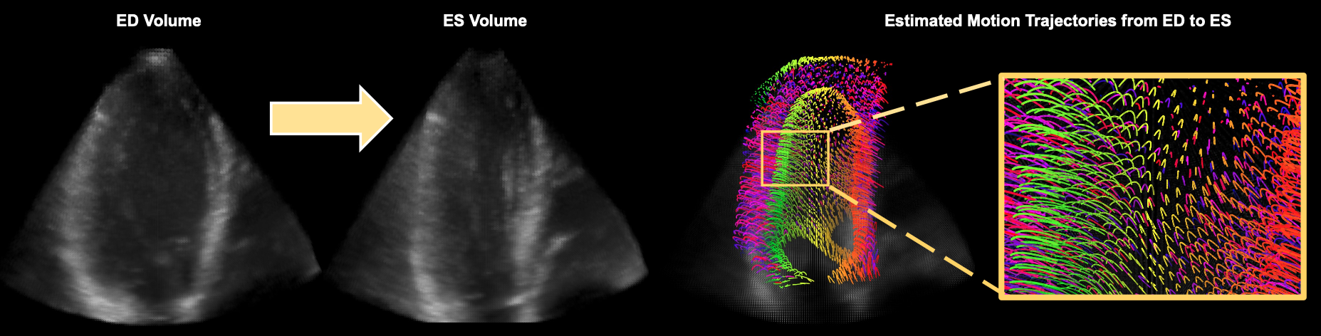
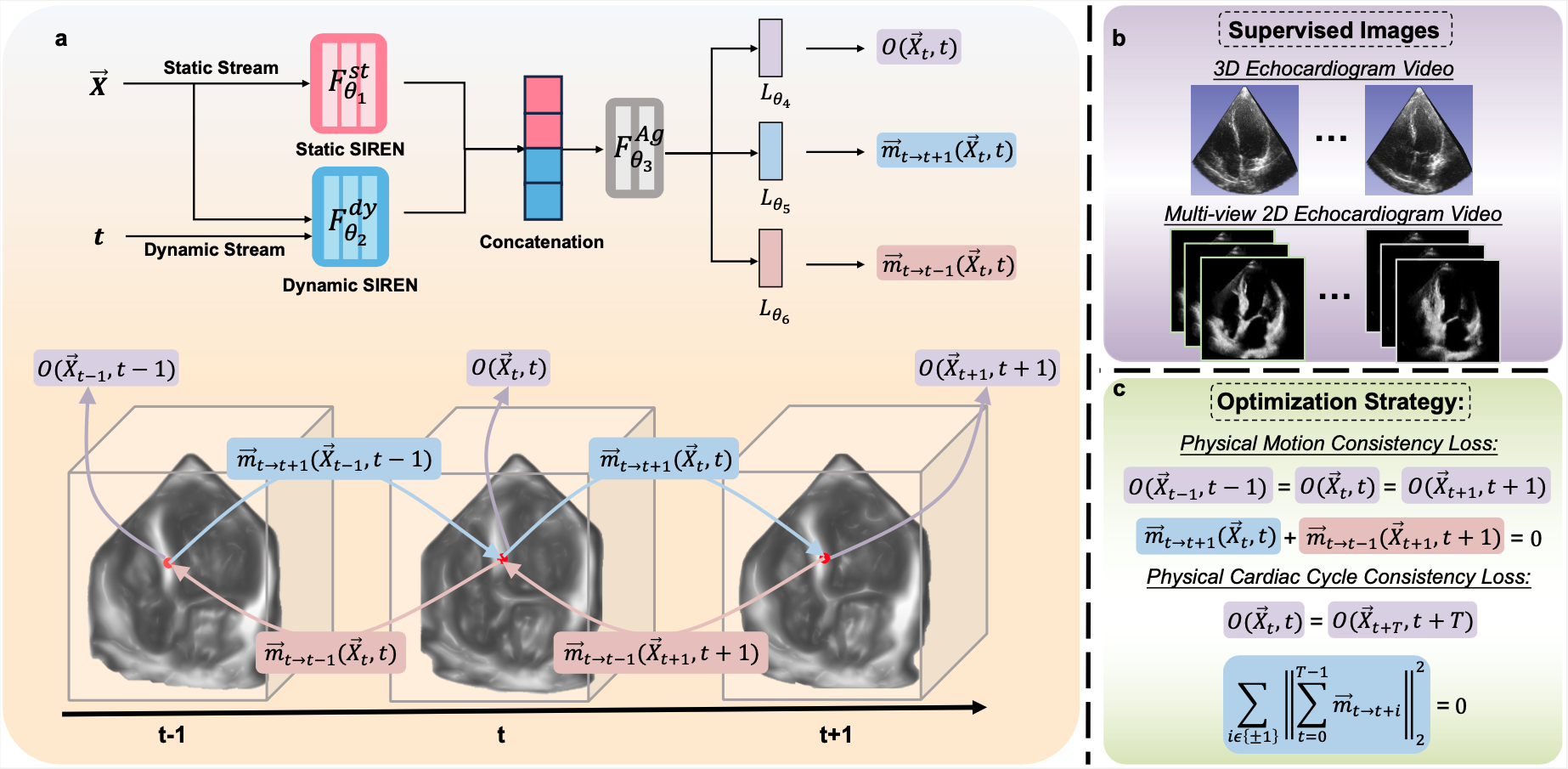
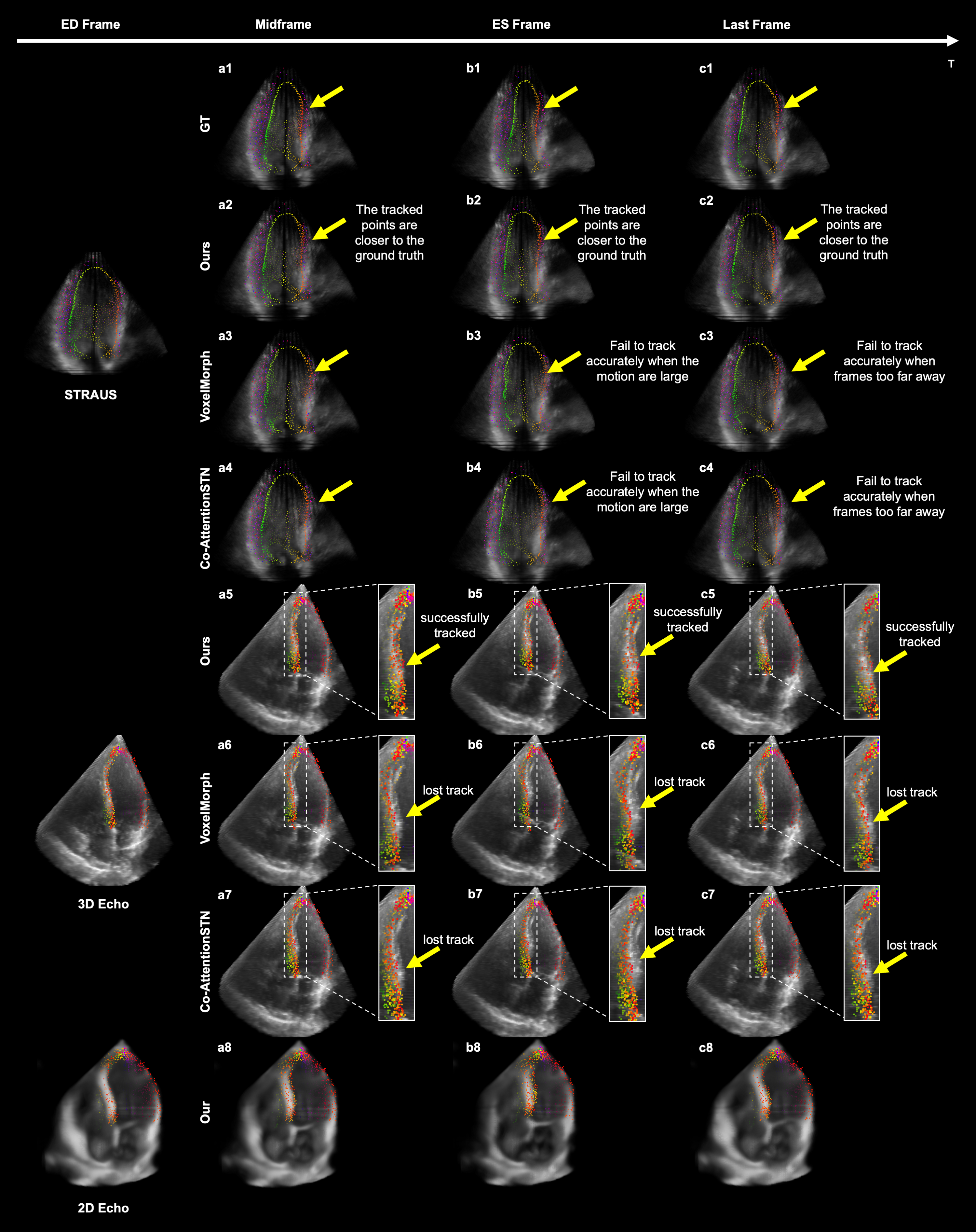
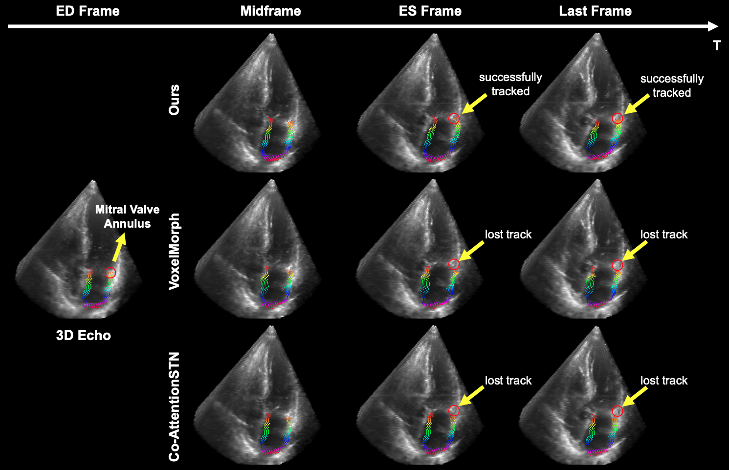
Myocardial motion tracking stands as an essential clinical tool in the prevention and detection of Cardiovascular Diseases (CVDs), the foremost cause of death globally. However, current techniques suffer incomplete and inaccurate motion estimation of the myocardium both in spatial and temporal dimensions, hindering the early identification of myocardial dysfunction. In addressing these challenges, this paper introduces the Neural Cardiac Motion Field (NeuralCMF). NeuralCMF leverages the implicit neural representation (INR) to model the 3D structure and the comprehensive 6D forward/backward motion of the heart. This method surpasses pixel-wise limitations by offering the capability to continuously query the precise shape and motion of the myocardium at any specific point throughout the cardiac cycle, enhancing the detailed analysis of cardiac dynamics beyond traditional speckle tracking. Notably, NeuralCMF operates without the need for paired datasets, and its optimization is self-supervised through the physics knowledge priors both in space and time dimensions, ensuring compatibility with both 2D and 3D echocardiogram video inputs. Experimental validations across three representative datasets support the robustness and innovative nature of the NeuralCMF, marking significant advantages over existing state-of-the-arts in cardiac imaging and motion tracking.

The workflow of our method

Description of the image
The figure illustrates the workflow of our method. (a) Inputs consist of 4D space-time coordinates (x,y,z,t). Corresponding outputs include the intensity value at specified space-time locations, as well as forward and backward 3D motion vectors that represent the movements of (x,y,z) at successive time frames t+1 and t-1, respectively. (b) Our approach can be applied to both 2D and 3D echocardiogram videos. (c) The methodology utilizes two main optimization strategies: physical motion consistency loss, ensuring that intensity remains constant within both the heart's tissues and blood-filled areas throughout the entire cardiac cycle, and physical cardiac cycle consistency loss, which aligns the process with the physiological fact that the heart muscle returns to its initial shape upon completing one full cardiac cycle.

Results

Point tracking results

Description of the image
This figure presents the qualitative results of point tracking using our method, VoxelMorph, and Co-AttentionSTN on the STRAUS, 2D, and 3D echocardiogram video datasets. The query points that need to be tracked during the end-diastole (ED) phase are displayed in the first column. The following three columns visually demonstrate the movement of these points across the full cardiac cycle. In the 2D echocardiogram video datasets, only our results are showcased since other methods are incompatible with 2D datasets.

Description of the image
This figure illustrates the tracking performance of our method, VoxelMorph, and Co-AttentionSTN for points on the left atrial (LA) myocardium within 3D echocardiogram video datasets. The initial column highlights the reference points set for tracking during the end-diastole (ED) phase. The subsequent three columns visually demonstrate the movement of these points across the full cardiac cycle. It is of significance to note that our method demonstrates superior accuracy in tracking points on the mitral valve annulus, a challenge not achieved by either VoxelMorph or Co-AttentionSTN.

4D heart resconstruction

BibTeX

@article{shen2023tracking,
  author    = {Shen, Chengkang and Zhu, Hao and Zhou, You and Liu, Yu and Yi, Si and Dong, Lili and Zhao, Weipeng and Brady, David and Cao, Xun and Ma, Zhan and Lin, Yi},
  title     = {Continuous 3D Myocardial Motion Tracking via Echocardiography},
  journal   = {arXiv preprint arXiv:2310.02792},
  year      = {2023},
}